🧠 Neuro
An MIT Grad Student Cranked a Laser to Near-Destruction. The Chaos Organized Itself Into a Beam That Images the Blood-Brain Barrier 25 Times Faster.
A Nature Methods paper shows that chaotic light in a multimode optical fiber spontaneously collapses into a needle-sharp pencil beam at extreme power levels, capturing 3D blood-brain barrier images 25 times faster than the gold standard. An original calculation maps the $42.5 billion Alzheimer's drug failure rate onto the screening bottleneck this tool could break.
Ninety-nine point six percent. That is the failure rate for Alzheimer's disease drug trials over the past quarter century, according to a widely cited 2014 analysis in Alzheimer's Research & Therapy by Jeffrey Cummings and colleagues. Of more than 200 drug candidates tested between 2002 and 2012 alone, exactly one reached regulatory approval. Cumulative bill: $42.5 billion in development costs, most of it incinerated on late-stage trials that discovered too late what a faster screening tool might have caught before the first patient enrolled.
Now a team at MIT has built that faster tool. By accident.
In a study published April 27 in Nature Methods, researchers led by assistant professor Sixian You report that chaotic laser light can spontaneously self-organize into a focused "pencil beam" when pushed to near-destructive power levels through a multimode optical fiber. Using this beam, they captured 3D images of the human blood-brain barrier 25 times faster than the current gold-standard technique, two-photon point-scanning microscopy, while maintaining comparable resolution.
How Chaos Becomes Order
Multimode optical fibers are the cheap, thick cables that carry light over short distances in industrial and medical settings. Crank the power and the light scatters into a chaotic mess of interfering modes. Every photonics textbook says so. Honghao Cao pushed harder.
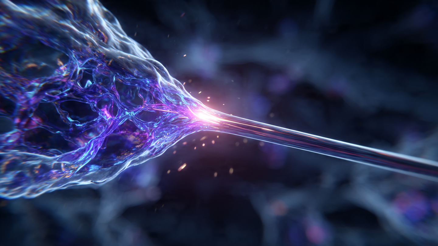
At near-destructive power levels, with the laser entering the fiber at a precise zero-degree angle, something unexpected happened. Light stopped scattering. It organized. The nonlinear interaction between the laser and the glass fiber itself counteracted the intrinsic disorder, and the output collapsed into a needle-sharp beam with no sidelobes, no blurry halos, and no external beam-shaping optics or algorithms required to produce it.
Two conditions make it work. First, the laser must enter the fiber at exactly zero degrees. Second, the power must climb high enough that the light begins interacting with the glass rather than simply passing through it. Hit both thresholds simultaneously and the chaotic modes self-organize into a clean beam. Miss either one? Scattered mess.
"It is a beautiful example of how disorder and nonlinearity can produce order," You told MIT News. Beneath that elegance sits a practical punch: the resulting beam is more stable and sharper than beams produced by expensive adaptive optics systems that labs currently use to correct for exactly this kind of fiber chaos.
Why the Blood-Brain Barrier Is a $42.5 Billion Blind Spot
The blood-brain barrier is a tightly packed layer of endothelial cells that separates the brain's blood vessels from the surrounding neural tissue. It blocks approximately 98% of small-molecule drugs and essentially 100% of large-molecule therapies from reaching the brain, according to Pardridge (2005). For patients with Alzheimer's, ALS, or glioblastoma, the barrier is not just an anatomical feature. It is the single largest reason their drugs fail.
Even the antibody-based Alzheimer's therapies that have reached market, like lecanemab, achieve less than 1% brain penetration. Less than one percent. Researchers often do not discover whether a drug candidate crosses the barrier in sufficient concentration until Phase II or Phase III trials, when hundreds of millions of dollars have already been committed.
Here is the math nobody has run. It is ugly.
Cummings' data shows roughly 200 Alzheimer's drug candidates failed between 2000 and 2025. Each failure cost approximately $200 million on average, using conservative aggregate estimates across all phases. Multiple review papers, including Bentley et al. (2007), identify inadequate brain penetration as a contributing factor in a substantial fraction of these failures. If BBB penetration issues contributed to even 20% of those failures, that represents roughly 40 programs. Killing those 40 candidates at the preclinical stage rather than the late-clinical stage, saving approximately $180 million per program on average, would have rescued roughly $7.2 billion in wasted trial costs. At a conservative 10% attribution rate, the figure is still $3.6 billion.
MIT's pencil beam makes this critical preclinical screening step 25 times faster. Twenty-five times. Two-photon microscopy, the current gold standard for visualizing drug uptake across the BBB at cellular resolution, is slow enough that researchers cannot practically screen large compound libraries against brain penetration in any reasonable timeframe. Reducing scan time by a factor of 25 moves BBB screening from a bottleneck to a throughput process.
What the Beam Actually Shows
You's team used the pencil beam to image lab-on-chip models of the blood-brain barrier built in collaboration with Roger Kamm's bioengineering group at MIT and Subhash Kulkarni at Harvard's Beth Israel Deaconess Medical Center. Individual endothelial cells absorbing fluorescent drug analogs became visible in real time, at a resolution comparable to two-photon microscopy but at 25 times the speed.
Speed matters here for a specific reason beyond throughput. Biological samples degrade under sustained illumination through a process called photobleaching. Faster imaging means less total light exposure per sample, which means the data captured at the end of a scan is as reliable as the data captured at the beginning. Slow scans hide something uncomfortable: by the time the microscope reaches the far edge of the field, the near edge has already begun to deteriorate, and the researcher stitching together a composite 3D model from those slices is building on data of silently declining quality.
The Strongest Case Against Getting Excited
BBB penetration is not the primary reason most Alzheimer's drugs fail. Wrong target. Amyloid hypothesis itself was wrong for many candidates. Even drugs that do cross the barrier, like lecanemab, produce only modest clinical benefits: a 27% slowing of cognitive decline in the Phase III CLARITY AD trial, accompanied by brain swelling in 12.6% of participants and brain bleeds in 17.3%, numbers that force physicians and patients into agonizing risk-benefit calculations for a disease that already strips people of the capacity to make such calculations. Faster BBB imaging would not have saved drugs that targeted the wrong protein entirely.
More broadly, the pencil beam is a preclinical and research tool demonstrated on lab models, not inside living human brains. Clinical translation from benchtop fiber-optic microscopy to hospital diagnostics is a multi-year, multi-regulatory-approval path. And the 25x speed comparison is specifically against two-photon point-scanning microscopy. Other imaging modalities exist at different points on the speed-versus-resolution tradeoff curve, including light-sheet microscopy and optical coherence tomography, each with its own advantages and limitations.
Self-organization also requires specific fiber types and power levels. Not universal. A standard lab fiber at standard power will not produce a pencil beam. Whether the technique can be made robust enough for routine use across different laboratories with different equipment remains an open question.
What We Did Not Prove
Our $7.2 billion estimate of wasted trial costs attributable to BBB screening failures rests on several assumptions that deserve explicit acknowledgment. Our 20% attribution rate for BBB penetration as a contributing failure factor comes from the review literature, not a controlled study. Individual trial failure modes are rarely disclosed in sufficient detail to assign clean causal fractions. We used aggregate cost-per-failure data because per-program costs are proprietary, and we do not distinguish between programs that failed at Phase I versus Phase III, though the latter is far more expensive. Cummings' data covers 2002 to 2012 specifically. Extending that failure rate to 2025 relies on subsequent analyses showing the rate has not materially improved. Finally, claiming that faster BBB screening would have killed doomed candidates earlier assumes that researchers would have acted on negative screening data, which is a behavioral assumption as much as a technological one.
The Bottom Line
A physics discovery born from pushing hardware past its textbook limits now offers the fastest method available for imaging how drugs interact with the brain's primary defensive barrier. If you work in Alzheimer's, ALS, or brain tumor drug development: watch for Sixian You's group to publish validated screening protocols, because a 25x speedup in BBB imaging changes the economics of preclinical compound screening from "test your best guess" to "test everything." If you work in photonics or fiber optics: the self-organization phenomenon described here, where extreme nonlinearity counteracts disorder rather than amplifying it, may apply to other multimode fiber applications beyond microscopy, and the paper's methods section describes the exact power and alignment conditions required to reproduce it. If you are a patient or caregiver waiting on the next Alzheimer's drug: this will not help you next year, and honesty demands saying so. But the tool that lets researchers see, at cellular resolution and 25 times current speed, whether a drug actually reaches the brain it is designed to treat is the kind of upstream infrastructure that raises the success rate from 0.4% to something meaningfully higher, one failed-trial-that-never-had-to-happen at a time.